
Services
Customized Retinal Solutions
Services offered by Dr. Lee
Expert Surgical Care for Complex Retinal Conditions
Our Services section is designed to help you feel informed and prepared throughout your care. Here, you can find helpful information about the diagnostic tests and procedures that Dr. Lee performs. We believe that clear education and communication are essential parts of high-quality retinal care, and our goal is to make your experience as smooth and comfortable as possible.
With extensive surgical experience, Dr. Lee provides advanced treatment for complex retinal conditions, including retinal laser, intravitreal injections, vitrectomy, scleral buckling, and secondary lens surgeries. Known for his meticulous technique and calm judgment, Dr. Lee delivers individualized treatment to ensure each patient receives precise care tailored to their unique conditions and needs.
Diagnostic Testing

Dilated Eye Exam
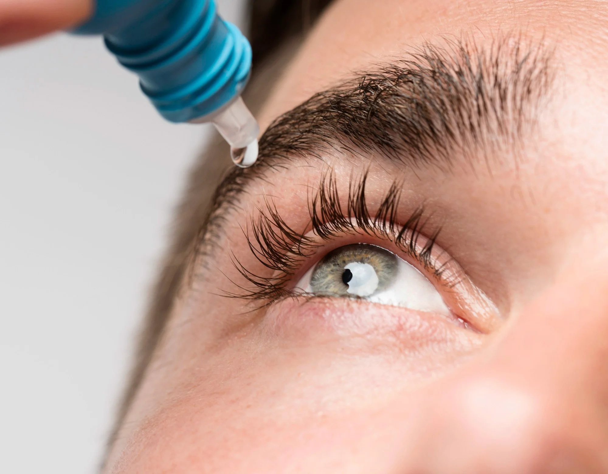
A dilated eye exam is a test in which a doctor uses special drops to widen the pupils so the entire retina can be examined. After the drops are placed, it usually takes about 15 to 30 minutes for the pupils to dilate. This exam is important for detecting and monitoring conditions such as diabetic retinopathy, macular degeneration, glaucoma, retinal tears or retinal detachment. During the exam, the bright lights can feel uncomfortable, and afterward vision may be blurry with increased sensitivity to light for 4-6 hours. Patients are often advised to bring their sunglasses to wear after the exam.
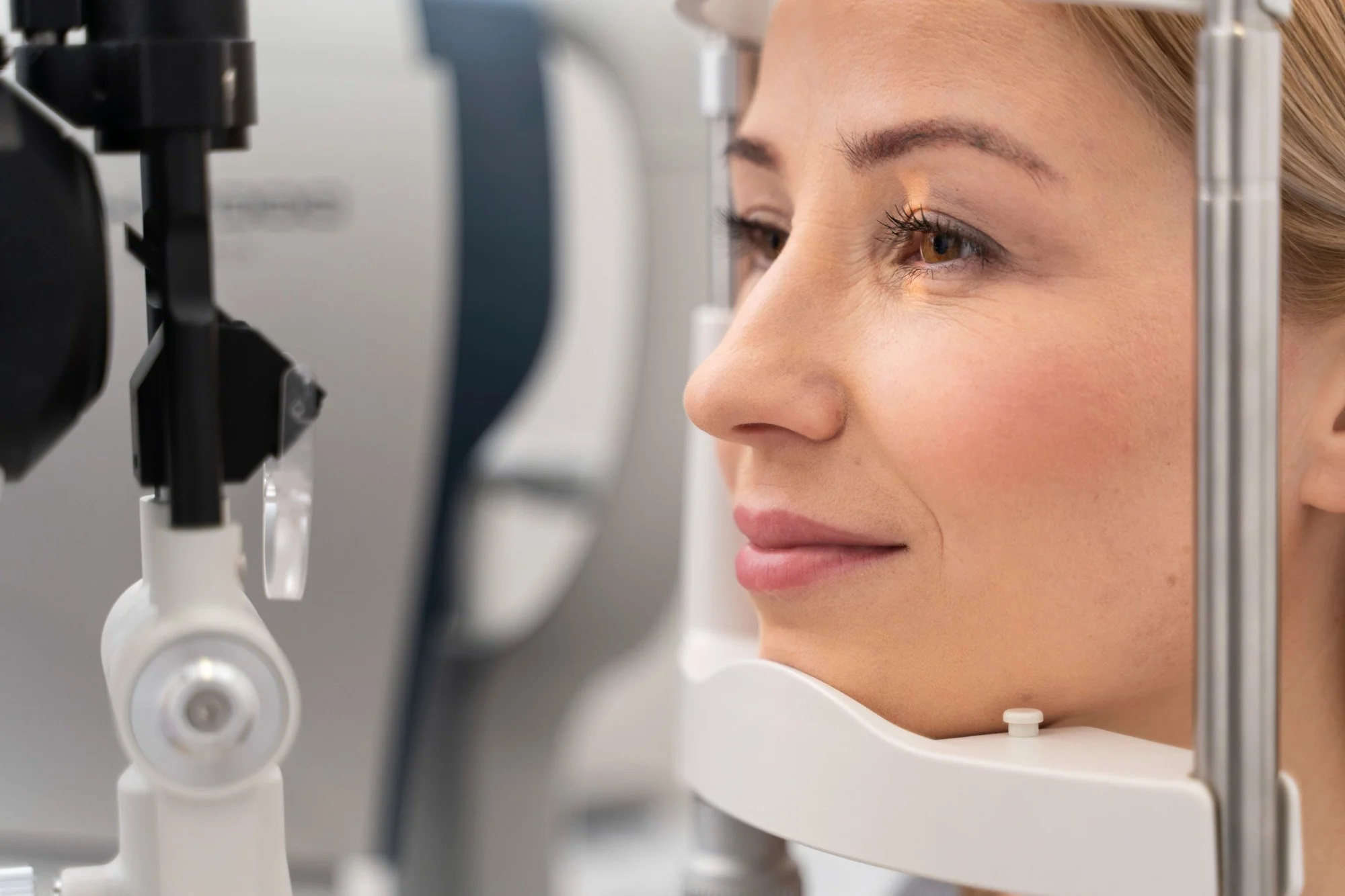

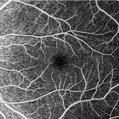
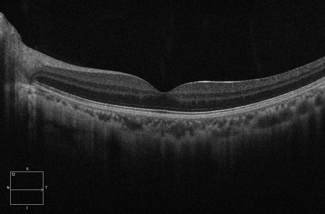
Retinal Imaging
-
A noninvasive imaging technique that uses light waves to produce high-resolution, cross-sectional images of the retina. It allows doctors to evaluate retinal layers, measure thickness, and detect subtle abnormalities such as retinal edema (swelling) or tissue damage.
-
An advanced imaging technique that captures high-resolution wide-field color images of the retina, optic nerve, and blood vessels. It is a valuable tool for diagnosing and monitoring retinal conditions like diabetic retinopathy, retinal tears, retinal detachments, and tumors.
-
A diagnostic eye test that uses a specialized camera and an intravenous dye (fluorescein) to visualize the blood circulation in the retina and choroid. This technique helps identify areas of blood vessel leakage, poor circulation, or abnormal new vessel growth.
-
A non-invasive imaging test that uses sound waves to create cross-sectional images of the eye when direct visualization of the retina is not possible, in cases of dense cataracts, vitreous hemorrhage, or ocular trauma. It helps detect retinal detachments, tumors, or other structural abnormalities.
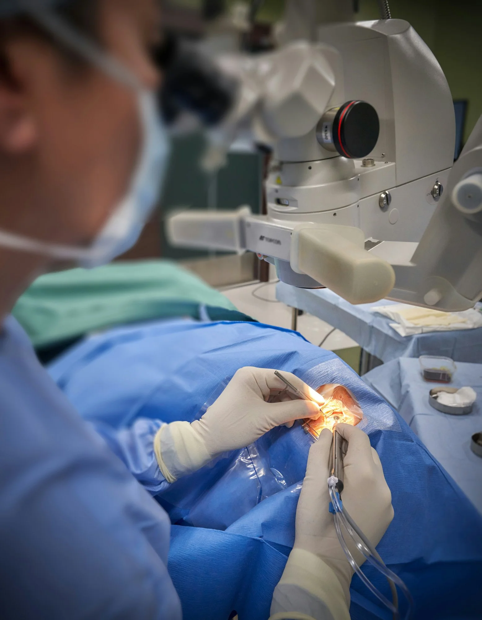
Procedures
-
Intravitreal Injection
An intravitreal injection is a medical procedure in which a doctor injects medication directly into the vitreous, the gel-like substance inside the eye. It is commonly used to treat conditions affecting the retina, such as age-related macular degeneration, diabetic retinopathy, retinal vein occlusion, and certain inflammatory or infectious eye diseases. It is performed in a clinic setting using local anesthetic and antiseptic solutions to minimize discomfort and reduce the risk of infection. Mild irritation or redness afterward is common, while serious complications are rare.
-

Laser Photocoagulation
Retina laser photocoagulation preserve vision by using focused light energy to seal leaking blood vessels, inhibit abnormal blood vessel growth, or reinforce areas of retinal weakness. The laser works by creating small, controlled burns that reduce fluid leakage, improve retinal oxygenation, and create a barrier layer to prevent retinal detachment. It prevents vision-threatening complications associated with conditions such as diabetic retinopathy, retinal vein occlusion, and retinal tears or holes.
-

Vitrectomy
Vitrectomy is a highly specialized microsurgical procedure in which the vitreous gel is removed from the inside of the eye to treat disorders affecting the retina and vitreous. Removal of the vitreous improves visualization and provides access to the retinal surface, allowing the surgeon to precisely repair underlying pathology. The vitreous is subsequently replaced with a balanced saline solution, intraocular gas, or silicone oil, depending on the clinical indication and surgical objectives. It is commonly indicated for conditions such as retinal detachment, vitreous hemorrhage, macular hole, epiretinal membrane, advanced diabetic retinopathy, as well as intraocular infections or ocular trauma. Vitrectomy is performed in an operating room under local or general anesthesia.
-

Scleral Buckle Surgery
Scleral buckle surgery is a procedure used to repair a retinal detachment by externally supporting the eyewall. During the procedure, the surgeon places a flexible silicone band or sponge along the sclera (the white outer layer of the eye), creating an inward indentation that relieves vitreoretinal traction and facilitates reattachment of the retina. It is combined with adjunctive retinopexy—either laser photocoagulation or cryotherapy—to seal retinal breaks. Scleral buckle surgery is performed in an operating room under local or general anesthesia.

Secondary intraocular lens surgery
Secondary intraocular lens surgery is a procedure performed to implant an artificial lens when a lens was not placed during a prior cataract surgery or when an existing intraocular lens needs to be replaced or repositioned. It is commonly done for patients who were left without a lens (aphakia), have a dislocated or unstable lens implant. This surgery is performed with vitrectomy, which facilitates safe placement and fixation of the intraocular lens while minimizing the risk of postoperative complications such as vitreous prolapse, retinal tears, or retinal detachment. It is performed in an operating room under local or general anesthesia.
Get in touch
Contact Dr. Lee today to book your appointment.